Morphologic verification of diagnosis is extremely important in oncology practice, because it is histologic diagnosis that allows for a very accurate differentiation of malignant tumors from other tumors. Immunohistochemical tests, currently used in labs, help determine the nature of cancer and choose the most effective ways of its treatment by determining the sensitivity of the tumor to a particular drug.
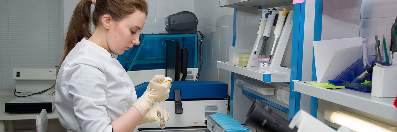
The main purpose of laboratory studies is morphologic diagnosis of diseases, which involves mainly microscopic research methods of pathologically altered tissues
Lab physicians study histology slides (glass) - thin sections of biopsy or surgery tissues stained with special dyes and placed between the slide and cover glass. Besides slides, tissue specimens may also be embedded in paraffin blocks – samples of pathologically altered tissue placed in paraffin – sliced later into very thin layers and examined under a microscope.
ATTENTION, very important: remember to pick up your glasses and histology blocks obtained during surgery or biopsy and store them properly! Only by having the glasses and blocks available you can always verify your diagnosis, conduct additional studies and develop an individual treatment plan.
Our lab provides the following services:
- Pathology consultation on available slides/blocks
- Consultation and rendering a second and third opinion of disputed morphology diagnoses from other health-providers in case of diagnostic discrepancies in the original morphology studies.
- Immunohistochemical studies
- Preparation of histological specimens
- Correction of available histological slides
- Histology studies of biopsy specimens
Our advantages
- Highly qualified professionals with years of experience - pathologists and lab technicians. The personnel of our laboratory actively participate in international and national research; attend all major oncology, neurosurgery and morphology meetings, conferences, congresses.
- MIBS laboratory is equipped with the cutting-edge equipment that allows for advanced histological and immunohistochemical techniques required for prompt morphology diagnosis in accordance with international standards. All of the lab equipment is manufactured by the industry leader - Leica Microsystems.
- Coordination among the specialists in chemotherapy, radiation diagnosis, radiosurgery, neurosurgery, and genetics. Collegial approach to patient management ensures the most accurate diagnosis, and as a result, selection of the most effective treatment strategy.
- Possibility to enlist the help of prominent Russian and foreign experts in morphology. Our laboratory cooperates with leading clinics in St. Petersburg and Russian Federation (Polenov Research Institute, St. Petersburg State University, Kirov Military Medical Academy and others), ensuring continuity in the patient management.
To get a complete and quality advice from our experts, you need to provide the following:
- Slides (available specimens)
- Blocks (tissue samples embedded in paraffin)
- Relevant medical records
- Copies of all the morphology (histology) reports issued at the previous stages of examination
- In case of head (spinal) tumors – pre-surgery MRI report
Studies completion time
- If no immunohistochemical tests are required and the submitted specimens do not need to be repaired, the report is normally provided in 1-2 days.
- If immunohistochemical tests are required, the study report will be provided in 3-7 days. In case of the second and third stages of immunohistochemical tests, the completion time may be extended.